<< back to Pathology Atlas menu
Pathology Atlas: Fallopian Tube
Normal Tube
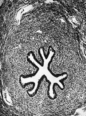 Photomicrograph showing the isthmic portion of the fallopian tube; it is
in this portion of the tube that spasm may occur and close the lumen. The
mucosa is lined by columnar epithelium which surrounds the lumen. The
columnar cells have cilia. The circular muscle layer is thickest
at the isthmus and thinnest at the infundibulum. Photomicrograph showing the isthmic portion of the fallopian tube; it is
in this portion of the tube that spasm may occur and close the lumen. The
mucosa is lined by columnar epithelium which surrounds the lumen. The
columnar cells have cilia. The circular muscle layer is thickest
at the isthmus and thinnest at the infundibulum.
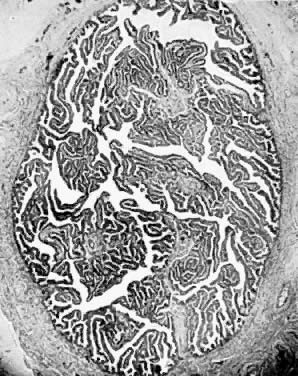 Photomicrograph (low power) of the human uterine tube. The mucosa forms
folds which in transsection of the tube simulate glandular structures. There
are, however, no true secreting glands in the oviduct. Photomicrograph (low power) of the human uterine tube. The mucosa forms
folds which in transsection of the tube simulate glandular structures. There
are, however, no true secreting glands in the oviduct.
Back to Top
Tuberculous Salpingitis
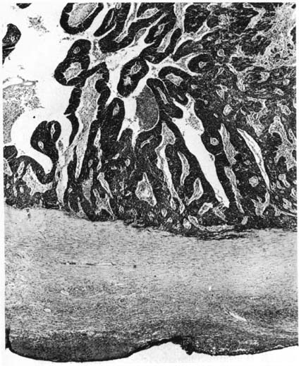
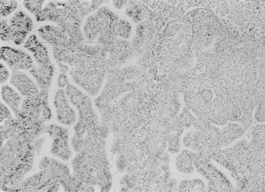 Tuberculous salpingitis. Chronic salpingitis due to tuberculosis presents
the characteristic histologic features of the tuberculous granuloma: lymphocytes, epithelioid
cell granulomata, and giant cells of both the
Langerhans and the foreign body type are seen. Tuberculous infection
of the fallopian tube often results in an “adenomatous” proliferation
of the lining epithelium. This is seen on the left of
this photomicrograph and may give rise to confusion with adenocarcinoma.(×100.) Tuberculous salpingitis. Chronic salpingitis due to tuberculosis presents
the characteristic histologic features of the tuberculous granuloma: lymphocytes, epithelioid
cell granulomata, and giant cells of both the
Langerhans and the foreign body type are seen. Tuberculous infection
of the fallopian tube often results in an “adenomatous” proliferation
of the lining epithelium. This is seen on the left of
this photomicrograph and may give rise to confusion with adenocarcinoma.(×100.)
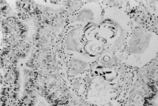 Tuberculous salpingitis may contain Schaumann bodies, which are more characteristic
of sarcoidosis than tuberculosis. These are conchoidal, laminated, calcified
structures, usually surrounded by foreign body giant
cells.(×100.). Tuberculous salpingitis may contain Schaumann bodies, which are more characteristic
of sarcoidosis than tuberculosis. These are conchoidal, laminated, calcified
structures, usually surrounded by foreign body giant
cells.(×100.).
Back to Top
Fallopian Tube Carcinoma
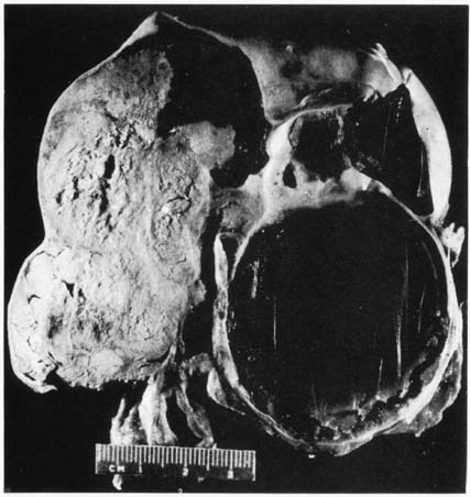 Primary carcinoma of the fallopian tube. The wall is smooth and grossly
uninvolved. The lumen is distended with tumor on the left and coagulated
fluid on the right, simulating a hydrosalpinx or pyosalpinx.(Courtesy of J. Donald Woodruff, MD) Primary carcinoma of the fallopian tube. The wall is smooth and grossly
uninvolved. The lumen is distended with tumor on the left and coagulated
fluid on the right, simulating a hydrosalpinx or pyosalpinx.(Courtesy of J. Donald Woodruff, MD)
 Papillary tumor with extensive growth into the lumen; the muscularis is
spared.(Courtesy of J. Donald Woodruff, MD) Papillary tumor with extensive growth into the lumen; the muscularis is
spared.(Courtesy of J. Donald Woodruff, MD)
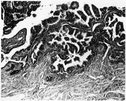 Papillary lesion developing from the normal epithelium as noted on the
left. Alveolar or pseudoadenomatous pattern is demonstrated on the right.(Courtesy of J. Donald Woodruff, MD) Papillary lesion developing from the normal epithelium as noted on the
left. Alveolar or pseudoadenomatous pattern is demonstrated on the right.(Courtesy of J. Donald Woodruff, MD)
Back to Top
Pelvic Inflammatory Disease (PID)
 Pelvic
Inflammatory Disease (PID) (From Operational Obstetrics
& Gynecology - 2nd Edition, The Health Care of Women in Military Settings,
CAPT Michael John Hughey, MC, USNR, NAVMEDPUB 6300-2C, Bureau of Medicine
and Surgery, Department of the Navy, 2300 E Street NW, Washington, D.C.
20372-5300, January 1, 2000. Original image courtesy CAPT Richard Stock,
MC, USN) Pelvic
Inflammatory Disease (PID) (From Operational Obstetrics
& Gynecology - 2nd Edition, The Health Care of Women in Military Settings,
CAPT Michael John Hughey, MC, USNR, NAVMEDPUB 6300-2C, Bureau of Medicine
and Surgery, Department of the Navy, 2300 E Street NW, Washington, D.C.
20372-5300, January 1, 2000. Original image courtesy CAPT Richard Stock,
MC, USN)
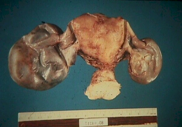 Pelvic
Inflammatory Disease (PID), Bilateral Hydrosalpinx
(From Operational Obstetrics & Gynecology - 2nd Edition, The Health
Care of Women in Military Settings, CAPT Michael John Hughey, MC, USNR,
NAVMEDPUB 6300-2C, Bureau of Medicine and Surgery, Department of the Navy,
2300 E Street NW, Washington, D.C. 20372-5300, January 1, 2000. Original
image courtesy CAPT Richard Stock, MC, USN) Pelvic
Inflammatory Disease (PID), Bilateral Hydrosalpinx
(From Operational Obstetrics & Gynecology - 2nd Edition, The Health
Care of Women in Military Settings, CAPT Michael John Hughey, MC, USNR,
NAVMEDPUB 6300-2C, Bureau of Medicine and Surgery, Department of the Navy,
2300 E Street NW, Washington, D.C. 20372-5300, January 1, 2000. Original
image courtesy CAPT Richard Stock, MC, USN)
Back to Top
Tubal Ectopic Pregnancy
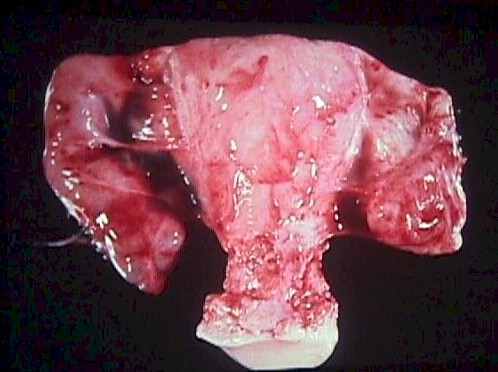
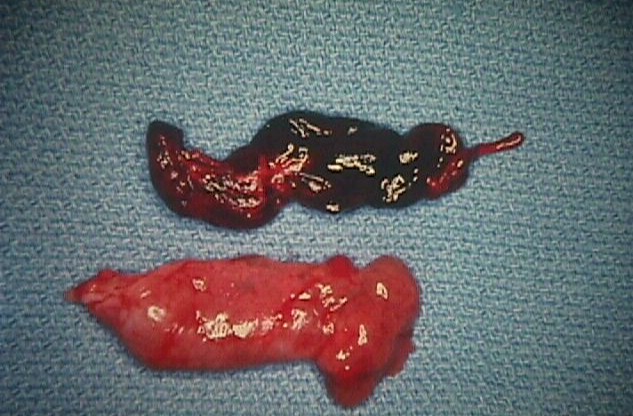 Tubal
ectopic pregnancy, with blood cast of the tube (From
Operational Obstetrics & Gynecology - 2nd Edition, The Health Care
of Women in Military Settings, CAPT Michael John Hughey, MC, USNR, NAVMEDPUB
6300-2C, Bureau of Medicine and Surgery, Department of the Navy, 2300
E Street NW, Washington, D.C. 20372-5300, January 1, 2000. Original image
courtesy CAPT Richard Stock, MC, USN) Tubal
ectopic pregnancy, with blood cast of the tube (From
Operational Obstetrics & Gynecology - 2nd Edition, The Health Care
of Women in Military Settings, CAPT Michael John Hughey, MC, USNR, NAVMEDPUB
6300-2C, Bureau of Medicine and Surgery, Department of the Navy, 2300
E Street NW, Washington, D.C. 20372-5300, January 1, 2000. Original image
courtesy CAPT Richard Stock, MC, USN)
Back to Top |